Structural Engineers Studying Skeletons
What is the difference between a bone and a building?
There isn’t one—at least according to Timothy Truster, an associate professor in the Department of Civil and Environmental Engineering (CEE) who leads the Computational Laboratory for the Mechanics of Interfaces (CLMI).
“In our group, we consider ‘structures’ to be anything composed of materials that support mechanical loads for long periods of time and during activity,” Truster explained. “That can be buildings and bridges, but also airplanes, cars, wind turbine blades, and even human and animal skeletons.”
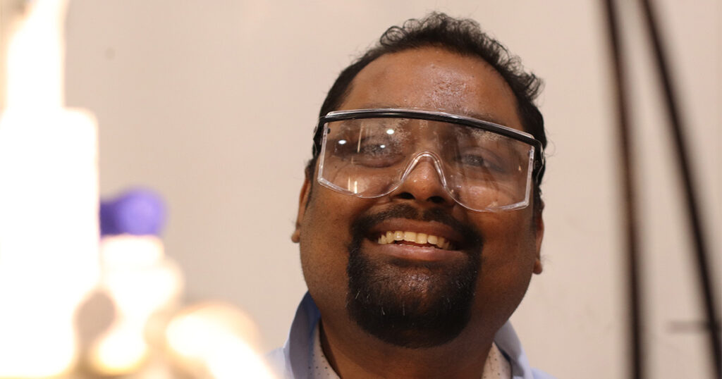
Members of the CLMI, including PhD student Debangshu Paul, apply mechanical theories to model the distributions of force and stress to predict and mitigate failures in various structures.
“While the fundamental principles of mechanics remain consistent, predicting long-term structural performance requires linking phenomena across vastly different scales,” said Paul, who specializes in computational mechanics. “This area bridges theory and practical engineering applications.”
Meanwhile, across campus, College of Veterinary Medicine Assistant Professor Pierre-Yves Mulon was bridging two areas of medicine. Many studies to further human health are performed in animals, but there are not many modeling programs that can accurately translate data between the two types of organism—so Mulon reached out to CLMI to create one.
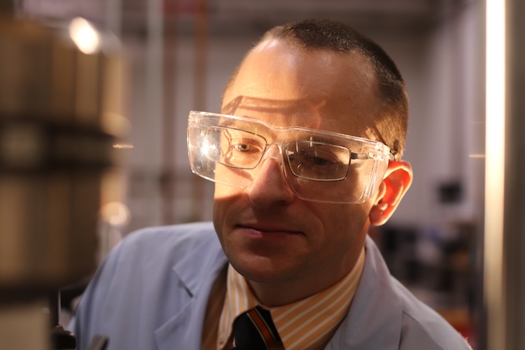
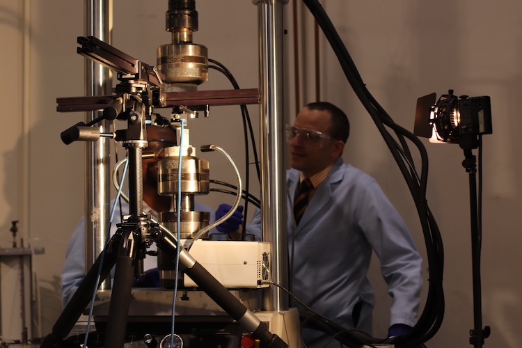
With Paul in the lead and funding from a University of Tennessee One Health Initiative seed grant, the team got to work. To validate their computational model against the behavior of actual goat tibia, Truster sought experimental support from CEE Fred N. Peebles Professor Dayakar Penumadu.
Earlier this year, Paul and his coauthors published their results in the open-access journal Biomedical Engineering Advances. It is the first published model of bone fractures that uses only free, open source software.
“The University of Tennessee system is well known for materials research as well as human and animal health research,” Truster said. “Together, our team’s clinical, simulation, and experimental expertise made it possible to take approaches from human patient-specific modeling and build this framework for goat tibia fracture prediction.”
Using Human Studies to Model Goat Bones
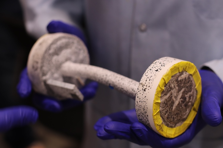
One of the most common bones to break—and the hardest to repair—is the tibia (shin bone). Due to the small amount of soft tissue protecting the bone, the risk of infection after a break is abnormally high. Since the bone bears a lot of the body’s weight, breaks frequently require orthopedic implants to reduce the chance of misalignment during healing.
“The tibia’s vulnerability to complications makes it a complex and critical site for studying and improving orthopedic implant performance,” explained Paul.
Scientists researching healing in human tibias commonly study tibia breaks and healing in goats, since the size and shape of the bones are similar. However, while there are several computational models for fracture and healing of human bones, there was no modeling framework for goat tibia.’
Paul’s team performed 3D quantitative computed tomography (QCT) scans of goat tibias, which were translated into a mesh simulation using open source software. The team then integrated techniques from studies on human bone structure and density to create a finite element (FE) model which would predict where and how goat tibias would crack under strain.
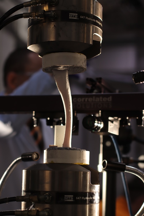
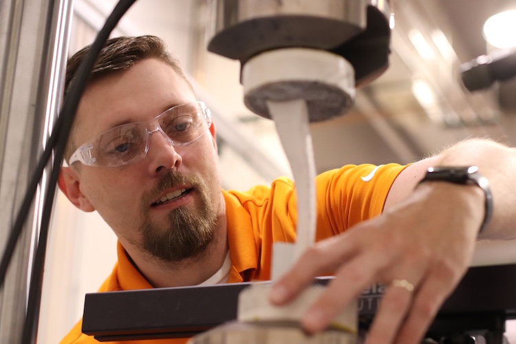
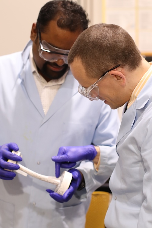
Paul went one step further, working alongside Penumadu’s PhD student Zachariah Arwood to experimentally strain the real bones and use digital image correlation (DIC) to directly compare the FE model’s results with real bone displacement and strain.
“Using DIC in this way gave us the unique capability to map and analyze prediction errors directly on the bone mesh, calculating and correcting for deviations in 3D,” said Paul.
Wide-Reaching Multidisciplinary Work
While the CLMI research philosophy is to see structural engineering concepts in unusual places, Paul said that getting to collaborate with medical experts was uniquely exciting.
“This project was a fantastic interdisciplinary experience,” he said. “It was exciting to learn from experts in different fields and to be exposed to new technologies throughout the process.”
To bring that multidisciplinary expertise to as many medical and structural researchers as possible, Paul and his coauthors were sure to base their model on free, open source software and publish their results in an open-access journal.
Paul hopes that the model will be adopted by many research teams who keep adding new capabilities. He is currently looking forward to modeling bone regeneration studies. Simulating goat tibias with defects or missing sections would improve those studies, helping translate their results more quickly back into human bones.
“Our framework makes it easier for scientists to study bone fractures and test new treatments—both in animals and, eventually, in people,” Paul said.
Contact
Izzie Gall (865-974-7203, [email protected])